USA


Home Blog Page 8

Poultry Market Intelligence Forum to Provide Industry Outlook and Food Systems Consumer Insights at IPPE

USPOULTRY will host its annual Poultry Market Intelligence Forum at the 2025 International Production & Processing Expo (IPPE) in Atlanta, Ga. This year’s forum will be held from 9 a.m. to 12 p.m. on Wednesday, Jan. 29, 2025, and is complimentary for all registered IPPE attendees.

The program will feature economic industry experts offering their insights on the current state of the global poultry industry and their forecasts for 2025. In addition, a food and nutrition expert will provide insight into identifying and addressing communication gaps related to food. The speakers will highlight key challenges facing the poultry industry, explore how the U.S. and international poultry sectors are positioned for the future, and address misinformation, while promoting science, to advance the food systems dialogue from farm to fork. The speakers and topics to be covered are as follows:

Economic Update
Mark Jordan, executive director
LEAP Markets Analytics

2025 Washington Update
Christian Richter, principal
The Policy Group

Going the Distance: Engaging from Farm to Fork
Wendy Reinhardt Kapsak, president and CEO
International Food Information Council

IPPE will offer a series of complimentary educational programs in addition to the Poultry Market Intelligence Forum. IPPE will take place at the Georgia World Congress Center. For more information and to register and reserve housing for the 2025 IPPE, visit ippexpo.org.

2025 IPPE SHOW DATES/HOURS:
Tuesday, Jan. 28: 10 a.m. – 5 p.m. EST
Wednesday, Jan. 29: 9 a.m. – 5 p.m. EST
Thursday, Jan. 30: 9 a.m. – 3 p.m. EST

Source: US Poultry & Egg Association

The Chicken or the Egg?

We may call ourselves the Peach State, but when it comes to raw numbers, poultry is unquestionably the king of Georgia agriculture.

And the chicken business is booming.

On an average day, Georgia farmers produce 30 million pounds of chicken, 7.8 million table eggs, and 6.5 million hatching eggs, contributing more than $25 billion to the state economy every year.

For decades, researchers in the University of Georgia’s Department of Poultry Science have worked at the forefront of the discipline, and their innovations in genetics, nutrition, and disease prevention have transformed the industry both at home and abroad.

Now, scientists working in this critical field will reach even greater heights thanks to the addition of a new state-of-the-art Poultry Science Building on UGA’s main campus. The $54.1 million, five-story structure comes equipped with more than 70,000 square feet of research and instruction space that will allow students,

Opening New Doors

The original Poultry Science Building opened during the Eisenhower administration on East Campus Road. It hosted one of the state’s most important departments for more than half a century, but the facility was not keeping pace with the work being done inside.

“Issues facing the poultry sector have grown inordinately complex. Faculty in our prior space were mostly within their own labs,” says Todd Applegate, chair of the Department of Poultry Science. “In our new building, we have clustered faculty into six thematic areas to face these current and future issues. Additionally, this environment allows our students a more robust experience garnering different research perspectives.”

Every classroom, student innovation space, and research lab in the new Poultry Science Building has been planned down to the individual chair. It includes massive, modular classrooms that give students comfortable spaces to learn, plus a basement facility where chicks and hens of all ages and sizes can be cared for. The walls are adorned with local, chic farm art, and the frosted doors feature environmental patterns that reflect the industry. That’s largely thanks to the support of donors and alumni, who had valuable experiences of their own in poultry science.

We wouldn’t be able to have a Chick-fil-A sandwich for under $9.50 if we didn’t make the progress that we do in our departments.” — Andrew Benson, associate professor in poultry science

Hands-on, innovative education is a cornerstone of the new building and the program it houses. For example, associate professor Andrew Benson BSA ’02, PhD ’06 incorporates lessons in anatomy and surgical demonstrations into his courses, lessons that would be difficult to teach in regular laboratories.

Benson aims to show students the real-world implications of their studies. His own work, which centers on fertility improvements, has allowed farmers to significantly increase the number of healthy chickens hatched and raised for the poultry industry.

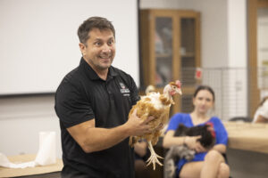
Associate professor Andrew Benson hosts “Hug a Chicken Day” for his Introduction to Poultry Science class. (Photo by Peter Frey/UGA)

“Every one of our labs is engaged with research that is addressing a need or opening up future endeavors for the poultry industry,” Benson says. “We wouldn’t be able to have a Chick-fil-A sandwich for under $9.50 if we didn’t make the progress that we do in our departments.”

Benson utilizes two different 1,500 square-foot learning labs lined with microscopes, incubators, and other scientific tools to help students participate in the research process. That’s just a few floors beneath two new, specialized research labs at the ready for students and faculty.

“I get to teach the course that I fell in love with, and that’s really why I like the new building the most,” he says. “Everything that I fell in love with about this department is still intact. Now, it just has a prettier face.”

A New Generation of Poultry Scientists

It’s not just the prestige of the new building that keeps poultry science alumni involved with the department.

Outreach specialist Sarah Beth Kersey (center) convenes with students (from left to right) Jalen Potter, Anna Thomas, Cooper Mattox, and Tianna Ramey in the foyer of the new Poultry Science Building. (Photo by Peter Frey/UGA)

Sarah Beth Kersey BSA ’24 was in eighth grade when she and her class toured UGA’s old poultry science building on a field trip.

“I fell in love with the department,” Kersey says. “After the open house, I was completely set on coming to UGA and studying poultry science. We are bringing that strong community forward, and the new building is the icing on top of the cake.”

After earning her bachelor’s degree in poultry science, Kersey wanted to give back by reaching out to the next generation. She’s doing just that as a communications and outreach specialist for the department while also pursuing a master’s degree in agricultural education in the College of Agricultural and Environmental Sciences.

“Education is the common goal for my job. Being able to apply what I learned in the classroom about poultry and then show how it works in daily life is really big. I think it adds a unique aspect of poultry science—learning, then helping people expand their knowledge,” she says.

Associate professor Brian Kiepper MS ’03, PhD ’07 knows something about building up future poultry scientists. As the teacher behind a very popular introductory course in the field, he instructs 350 students every semester.

“My whole focus is for our students to understand how agriculture changed it all,” says Kiepper. “When they leave the University of Georgia as a doctor, lawyer, teacher, engineer, or whatever, they can be that because of agriculture. They couldn’t if somebody else didn’t take on the responsibility of creating those calories you need to survive.”

Kiepper can be creative in his teaching. For instance, in his First-Year Odyssey Seminar, “Chicken Que: Science Behind the Grill,” students prepare, flavor, and cook their own meals from scratch to truly understand the farm-to-table perspective.

It’s a creative way to learn about such an important industry beyond just numbers and slides. With the minds of the poultry science department and the capabilities of the new building, Georgia’s poultry leadership is well positioned to grow.

“When you see our new building, it really is a place to show people what we do,” Kiepper says. “With the old building, we had to entice people into poultry science. Now, with the new building, it’s almost like it makes that stamp itself.”

Abit Massey – A Lifetime of Leadership: A key component of the new poultry science building is the Abit Massey Classroom. Massey BBA ’49, who died in June, was a poultry industry giant, a beloved entrepreneur, and a strong UGA supporter. Following his graduation from the Terry College of Business, Massey’s tenure as executive director of the Georgia Poultry Federation made Georgia the poultry leader it is today. The classroom, adorned with a large display and official lettering, is dedicated to his memory, and it recognizes his role in making the university and the state a leader in the poultry sector. (Photo by Andrew Davis Tucker/UGA)

Source: The University of Georgia

Deadly Listeria Outbreak Sparks Nationwide Recall of Yu Shang Food Products

A listeria outbreak connected to ready-to-eat meat and poultry items from Yu Shang Food Inc. has led to 11 confirmed illnesses across four states, including nine hospitalizations and the tragic death of an infant, according to the U.S. Centers for Disease Control and Prevention (CDC).

Yu Shang Food, based in Spartanburg, South Carolina, is recalling over 72,000 pounds of its products due to potential contamination with Listeria monocytogenes. The recall covers all ready-to-eat meat and poultry products manufactured before October 28. These items were distributed to retailers nationwide and sold online, the U.S. Department of Agriculture’s Food Safety and Inspection Service (FSIS) reported.

The contamination was identified during routine testing by the FSIS last month. Genetic sequencing is ongoing to determine if the bacteria in the recalled products matches the strain responsible for the outbreak.

So far, the CDC has recorded seven cases in California, two in Illinois, and one each in New York and New Jersey. Investigators interviewed eight affected individuals, seven of whom reported shopping at markets where Yu Shang products were sold. Two specifically recalled consuming Yu Shang’s ready-to-eat chicken.

The CDC shared a heartbreaking update from California, where a pregnant woman and her twins became ill. Unfortunately, both infants passed away. While listeria bacteria were detected in the mother and one of the twins, no bacteria were found in the second twin. Therefore, only the mother and one twin are counted among confirmed cases. Another infant was also infected but has since recovered.

In a statement to CNN, Yu Shang Food acknowledged the woman’s report of consuming their products but emphasized that no direct evidence links their items to the illnesses.

Health officials caution that the actual number of cases may be underreported, as some individuals recover without seeking medical care. Moreover, linking illnesses to outbreaks often takes several weeks.

Consumers are urged to discard or return any recalled products immediately and to clean surfaces or containers that may have been in contact with the food. Listeria can survive in refrigerated environments, making proper cleaning critical.

Listeriosis, caused by Listeria monocytogenes, ranks as the third leading cause of death from foodborne illnesses in the U.S. Symptoms include fever, muscle aches, fatigue, and sometimes severe complications like stiff neck, confusion, or seizures. Those most vulnerable are older adults, pregnant women, and individuals with weakened immune systems.

Anyone who develops symptoms after consuming the affected products should seek medical attention promptly.

Source: CNN

Letter to the Editor: Workplace injury numbers in poultry industry need closer look

As FSN noted recently, the Bureau of Labor Statistics reported that poultry slaughter plants had a huge and unprecedented reduction in the number of workers injured this year: the industry injury rates were more than halved from the year before.  The BLS data also showed that poultry plants reduced their most serious work injuries that involve lost time or restricted duty by more than two thirds in just one year. What the FSN story didn’t report was that these numbers are based on data collected and reported by the industry, and the data are not checked or validated. Though the FSN story led with a celebration of these numbers, there are very serious questions about these self- reported numbers and whether they are real or a mirage.

Over the past decade three government agencies (USDAOSHA and NIOSH) have found that the poultry industry’s self-reported injury numbers are seriously underreported. Congressional investigations also documented this in a 2021 Congressional Committee report that found that the poultry industry flagrantly underreported the number of their workers sick with COVID-19 by two thirds.

Many of us were startled to see this one year unprecedented decrease in self-reported cases by the industry. For those of us that follow worker safety in the industry, we didn’t hear from workers about any new improvements or changes that would lead to this drop.  The last published studies of repetitive trauma injuries, like carpal tunnel syndrome, in poultry plants were performed by the National Institute for Occupational Safety and Health (NIOSH) in2012 and 2014.  NIOSH performed medical exams and found alarmingly high rates of carpal tunnel syndrome among line workers — rates from 34 percent to 42 percent.

Taking a closer look at these BLS numbers may help explain why they may be a mirage: BLS numbers are calculated from a company’s self-reported injury logs that contain only those injuries/illnesses that the company deemed were work related and where the worker received medical treatment from a doctor.  If the plant does not send workers to a doctor for treatment, they would have very few cases on their logs.

In poultry plants, government investigations found that injured and ill workers were seen in onsite health units and rarely referred to a doctor for medical evaluation and treatment.  The onsite clinic staff only provided first aid to the injured worker—not ‘medical treatment’ that would be considered a ‘recordable’ injury.

A  recent article in the American Medical Association’s Journal of Ethics documented myriad government investigations in big poultry plants that found that the onsite health clinics routinely send injured and ill  workers back to  jobs that cause their injuries,  instead of sending workers to a doctor for a diagnosis and treatment. In some cases, workers went to the onsite clinics dozens of times with the same injury, never to be sent to a doctor.  In these cases, effective treatment was delayed, and workers’ injuries worsened leading to surgery and other bad outcomes.

When a worker’s condition worsens and they then go to see a doctor on their own, the companies claim these injuries are not work related. The companies don’t pay for the medical treatment, and the injuries are not recorded. Studies and investigations also found that workers may be intimidated into not reporting work related injuries and illnesses for fear of losing their jobs.

The high turnover in the poultry industry, between 60 percent to 150 percent a year,  is often thought to be a consequence of injured workers who can no longer do the job, don’t get to be seen by a doctor to get adequate diagnosis and  medical treatment to recover —and must leave the industry. Of course, these injuries are also not recorded on the companies’ logs.

A closer look behind these numbers is warranted. What incredible improvements implemented last year have led to this drop? Are these low recordable injury rates really a reflection that the companies are much safer?

Source: Food Safety News

$7,000 USPOULTRY Foundation Student Recruiting Grant Awarded to Hannibal-LaGrange University

The USPOULTRY Foundation awarded a $7,000 student recruiting grant to Hannibal-LaGrange University (HLGU) in Hannibal, Mo. Through its bachelor’s degree in business administration with a concentration in agribusiness, HLGU equips students with a solid foundation in business principles and industry-specific knowledge, preparing them for successful careers in the poultry industry and other related fields.

The agribusiness program features a small demonstration farm on campus, which currently houses 42 chickens as the foundation of an egg production operation. This fall, the program will expand to include egg incubation and the beginning stages of raising meat chickens. This hands-on experience is complemented by a required agribusiness internship, allowing students to apply their knowledge in real-world settings. Additionally, poultry-related content is integrated into the Introduction to Agriculture course, providing students with a well-rounded foundation in agricultural practices.

The funds granted to HLGU will support targeted recruitment efforts aimed at regional high schools and FFA groups, helping to inspire young people and raise awareness about the benefits of poultry production and agribusiness. These funds will be used to create promotional videos for digital marketing campaigns as well as to design and produce flyers and giveaway items that will be distributed at regional recruitment events.

The USPOULTRY Foundation board approved student recruiting grants totaling $324,215 to 28 colleges and universities across the U.S. with a poultry science department or industry-related degree program. The Foundation provides annual recruiting and retention funds to colleges and universities to attract or connect students to their poultry programs or industry. The grants were made possible in part by gifts to the USPOULTRY Foundation from companies, individuals and families, in addition to funds earned over the years from the International Poultry Expo, part of the International Production & Processing Expo.

Source: US Poultry & Egg Association

All in One Basket – 11.22.24

This is a USPOULTRY podcast highlighting news and events affecting our membership and the feather industry.

Effect of breeder age and early hypoxic stimulation of the chorioallantoic membrane on vascularization, internal organ development, blood profile and chick organ histology

Breeder age significantly influences egg quality traits and subsequent embryonic development (Machado et al., 2020; Nasri et al., 2020; Zita et al., 2022). The composition of eggs at oviposition and oxygen concentration (O2) during incubation are crucial factors affecting nutrient metabolism during avian embryo development (Wangensteen and Rahn, 1970; Wilson, 1997). Despite advancements in artificial incubation, aspects related to the gaseous environment are still under investigation to understand epigenetic effects and enhance process efficiency (Decuypere and Bruggeman, 2007; Druyan et al., 2018; Okur et al., 2022). Oxygen supports embryonic growth through yolk beta-oxidation and conductance through shell pores, notably in older breeders (Decuypere and Bruggeman, 2007; Druyan et al., 2018; Okur et al., 2022). Gas exchange rates vary during different incubation stages, influencing embryo development and organ formation, each with distinct critical windows (Burggren and Elmonoufy, 2017).
The influence of O2 and carbon dioxide (CO2) on embryonic development, particularly hypoxia, a condition characterized by a deficiency in oxygen supply to tissues, has significant effects on internal organ development (Decuypere and Bruggeman, 2007). Hypoxia regulates angiogenesis, influencing embryo metabolism, growth, and health (Carmeliet, 2003; Verhoelst et al., 2011). Altimiras and Phu (2000) and Sharma et al. (2006) emphasize the importance of a specific threshold of O2 and its availability for initiating and sustaining early embryo development. Each hypoxic exposure or stimulation window tends to have an impact on embryo and organ growth and development. While mild hypoxia can improve gaseous diffusion capacity and mitigate detrimental effects on embryonic development, acute or sustained hypoxia during early development may negatively impact vital organ growth (Zhang and Burggren, 2012). However, a study suggested that chronic hypoxia during specific incubation periods may not affect embryo or organ weight at hatching (Miller et al., 2002). Hypoxia-induced changes and adaptations in embryos depend on the timing, intensity, and duration of exposure (Chan and Burggren, 2005; Stenmark et al., 2006; Storz et al., 2010; Burggren and Elmonoufy, 2017; Zhang et al., 2017; Storz and Cheviron, 2021). A variety of organs are affected by hypoxic conditions due to alterations in gene expression and physiological responses (Miot et al., 2012).
Hypoxic stimulation of the chorioallantoic membrane (CAM) is proposed to improve respiratory and cardiovascular development in embryos (Haron et al., 2021). Researchers have found that exposure to hypoxic environments can affect hemoglobin levels, indicating a physiological response (Huang et al., 2017). During development, this alteration in blood profile may be an adaptive response to reduced oxygen availability. Hypoxia also plays a role in the vascularization of the heart via its vasodilatory effects, once the coronary circulation is functional (Tomanek et al., 2003). This response is in line with the chick’s body’s attempt to increase oxygen supply or provide rapid support for angiogenesis under conditions of tissue damage especially to tissues of the heart and lungs, when reduced oxygen availability is present (Hsia et al., 2013). Pearce (2006) and Miller and Zachary (2017) stated that low oxygen levels may compensate for breeder aging, resulting in a more controlled and normal neovascular response in the heart and lung tissues of the chicks.
Hypoxia selectively affects Chorioallantoic membrane (CAM) development (Azzam and Mortola, 2007). Early-stage hypoxia affects the development of the CAM, enhancing its growth and vascularization (Druyan et al., 2012; Druyan and Levi, 2012; Druyan and Levi, 2012; Haron et al., 2021). Conflicting reports exist regarding CAM vascularization under hypoxia, with some studies showing no change (Burton and Palmer, 1992) or decreased effects (Burton and Palmer, 1992; Wagner-Amos and Seymour, 2003). While literature exists on these differences mostly for broilers, there is limited information on the effect on layer breeders, in terms of its breed and age. The developmental trajectory of embryos in broilers and layers is different in response to differences in gaseous exchange conditions during incubation. This research, therefore, investigates the effect of early hypoxic stimulation of two (2) layer breeder age eggs during embryogenesis on organ development, CAM vascularization, organ tissue histology and blood indices of chicks hatched as a consideration to adaptability to low oxygen during incubation.

MATERIALS AND METHODS

Experimental Site, Ethics and Facilities

This research was conducted at the University of Lomé at the Regional Center of Excellence for Poultry Science (CERSA-UL) hatchery, research farm and laboratory. All experimental procedures were approved by the Animal Ethics and Scientific Committee following the guidelines of the University of Lomé, CERSA (008/2021/BC-BPA/FDS-UL). The incubators used for the experiment were located at latitude 6°1′95′′N and longitude 1°2′53′′E with an elevation of 26 m above sea level (Google. n.d., 2024).

Experimental Design

A total of 900 eggs were tested in a 2 × 3 factorial arrangement of 2 breeder flocks ages (33- and -50 wk) and 3 oxygen concentration (O2) levels that include;

  • 1.
    15%, 17% O2 (experimental groups) and
  • 2.
    21% O2 (control group).
Each breeder age group had 450 eggs and in each group of O2 levels, 150 eggs were divided into 3 replicates of 50 eggs.
From embryonic day (ED) 7-9, a steady stream of air-N2 mixture was used to flush the experimental incubators for only 1 hr/d to reduce the O2 levels to 15% and 17% for the experimental groups. An O2 gas detector (Model HFP-1201 BX, No. D6924, Xi’an Huafan Technology Co., Ltd.) was used to continuously monitor the O2 levels (Druyan et al., 2012; Zhang and Burggren, 2012) in the three (3) PAS REFORM (PasReform, Zeddam, Netherlands, SmartPro Combi model) incubators used. The experimental group incubators were returned to equivalent incubation condition as the control after the 1 h exposure period each day.

Hatching eggs, storage and incubation conditions

The hatching eggs from ISA Brown layer breeders were collected and stored at 18°C temperature (T) and 75% relative humidity (RH) for 4 d. Following storage, the eggs were prewarmed at 24°C for 6 h before incubated. At egg setting, all groups of randomized hatching eggs were incubated in three (3) incubators calibrated to equivalent temperature of 37.7°C, relative humidty of 56% and automated turning at a 90° angle every hour. On the 3rd hour of each day of embryo age (ED 7, 8 and 9) from the time of setting, the two (2) experimental group incubators were continuously flushed with air-N2 for only 1 h. During the period of flushing, oxygen concentration was reduced in the various experimental incubators to 15% and 17%. The incubator’s O2 levels were continuously monitored and adjusted using the oxygen gas sensor. The experimental incubators were returned to calibrated equivalent condition as the control group after the 1 h oxygen reduction period. On d 18, eggs were candled to identify living embryos, which were then moved to hatching baskets for the 3-d hatching process.

Embryo and Chick Internal Organ Measurements

Following the ED 7-9 exposure to lowered oxygen concentrations, 6 embryos were sampled from each replicate at ED 11. Eggs were weighed and eggshells were broken in the airspace region for the embryo to be removed and weighed. The embryo was dissected for the weight of the heart and liver using a sensitive weighing scale (Ohaus STX8200 Scout). Weights obtained were used to calculate relative organ weights using the following equation:

At hatch, all chicks were weighed from each treatment group (data not shown). Nine (9) chicks from each treatment were humanely sacrificed by cervical dislocation and the fresh weight of the yolk sac, heart, liver and lungs was taken. Yolk-free chick weight was then estimated and used to calculate the relative organ weights using the equation:

Chorioallantoic Membrane Vessel Measurements

On ED 16, 1-2 mL of 10% formalin was injected through the airspace into three (3) eggs from each treatment group for retention of erythrocytes into vessels of the CAM. After 24 h, the egg was cut into 2 longitudinal sections along the equatorial region using a Dremel drill with a round blade. The embryo, yolk and albumen were carefully removed from the eggshell with little disturbance to the adhered CAM on the eggshell. A beam of 100W halogen light in an all-closed box-like device with a conical opening with a diameter 3 cm on 1 side was passed through the eggshell from the outer part. Digital images were taken with a Canon (EOS Rebel T5i) camera with a focused lens (EF-S 18-55 mm) at 2 different spots in each half section of the longitudinally divided eggshell. A spot represents the circular region covered by the beam of light that passes through the conical-sided opening of the box. From each circular spot image, 2 regions of interest (ROI), each 400 × 400 pixels were cropped for analysis. The ROI is the subset of the spotted image that was manually identified based on clear vessel branches. This procedure followed slight modification from Verhoelst et al. (2011) and Fernandes et al. (2017).
Eight cropped ROI images were obtained from each egg. A total of 24 images/treatment group were processed and analysed as described by Fernandes et al. (2017), using Image J software (version 1.54j, NIH, USA) for the vascular fraction (VF) (%), and fractal dimension (FD) of the vascular network of the CAM (Figure 1). The FD is the measure of the degree of branching of vascular network while the VF is determined as a measure of vessel density, which is in percentage (%) (Verhoelst et al., 2011; Fernandes et al., 2017).
Figure 1

  1. Download: Download high-res image (662KB)
  2. Download: Download full-size image

Figure 1. Image processing procedure using Image J software. Abbreviation: ROI, region of interest: The region of interest (ROI) is the subset area of the main initial image cropped to 400 × 400 pixels for analysis. The region was manually chosen based on clearly defined major and minor vessel branching.

Blood Sampling and Analysis

At hatch, blood samples were collected from the heart of nine (9) chicks from each group with a 27-G needle and 1 mL syringe into ethylene-diamine tetra-acetic acid (EDTA) and plain gel tubes. The blood samples in the EDTA tubes were promptly analyzed using an automated hematoanalyzer (DH36, Dymind Biotechnology) for haematological parameters while blood stored in the gel tubes were centrifuged at 3000 rpm at 15 min to obtain serum samples which were stored at -20°C until Triiodothyronine (T3), thyroxine (T4) and biochemical analysis. A volume of 100 mL of serum was used for T4 and T3 concentration determination in an automated VIDAS system, which is an enzyme-linked fluorescent assay (ELFA) technique. The antibodies, anti-T3 and anti-T4 of mutton, provided by VIDAS were used in the assay for the determination of the concentrations of T3 and T4, respectively. Serum biochemistry was analyzed by the colorimetric method using an automatic device for total protein (TP), albumin (ALB), total cholesterol (TC), triglycerides (TG) and high-density lipoprotein (HDL) cholesterol. low-density lipoprotein (LDL) cholesterol was estimated using the formula; LDL-chol = [TC] – [HDL-chol] – [TG]/5.

Histological Analysis

Heart, lung and liver tissues dissected from the set of chicks were fixed in 10% buffered formalin. Thick sections of 5 µm were cut from the paraffin-embedded blocks after a series of alcohol (100, 96, 80, and 70%) and xylol treatments, deparaffinized in xylol and stained with Hematoxylin and Eosin. A 100um microphotograph image was taken under a light microscope (Thermo Fisher Scientific, Massachusetts) after examination. As used by Okur et al. (2022), a similar grading score was emplored by a histhopathogist in grading the neo-vascularization in heart, lungs and liver tissues. The neo-vascularization grading score include; ++++ = Very high (ectatic vessels with high congestion); +++ = high (vessels seat of moderate congestion); ++ = Normal (normal tissues).

Statistical Analysis

The data were processed with the statistical software package GraphPad Prism 6.0 (GraphPad Software, La Jolla, CA). The factors: breeder age (Ab) and oxygen concentration (O2) levels were used in a 2 × 3 factorial arrangement. The statistical analyses of the results were performed using the 2-way analysis of variance (ANOVA) model. All data obtained for the analysis were first transformed using the arc sinus data transformation rule and then tested using the Shapiro-Wilk test for normality (Levene’s test for homogeneity of variance). The 2-way ANOVA designs used followed the general linear model (GLM) procedure which is as follows:
WhereYijk is the Dependent Variable
µ is the overall mean,
Ai is the effect of age (i= 33- and -50 wk),
O2j is the effect of the O2 levels (j = 15%, 17% and 21%) in the experiment,
AO2ij is the effect of the interaction between Ab and O2, and
eijk is the random error term.
The post-hoc Tukey test was used to separate and compare the means of each parameter in relation to the effect on Ab, O2 level and the interaction (Ab * O2) between the 2 factors. Means were compared and separated at a significant level of 5% (P < 0.05).

RESULTS AND DISCUSSION

Relative Organ Weights

Table 1 summarizes the impact of breeder age (Ab) and oxygen concentration (O2) level on embryo and chick organ weight taken at ED 11 afterexposure and d 1 respectively. The relative weight of organs of the embryo was with regards to embryo weight while those of the chicks were with regards to yolk-free chick weight (data not shown for embryo weight and yolk-free chick weight). No significant (P > 0.05) changes were noted in relative heart weight at ED 11, but the relative liver weight was affected by O2 levels, with the 21% O2 group having significantly (P = 0.003) lower weight than low O2 levels. Interactively, chicks from the 50 wk breeders incubated at 21% O2 level exhibit superior relative heart weight (P < 0.001) compared to 15% and 17% low O2 levels of the same breeder age. Additionally, the 33 wk breeders had relatively higher heart weights (P = 0.013) than the 50 wk breeders. The main effect of O2 levels on relative heart weight reveals significantly higher weight (P < 0.001) at 21% O2 compared to 15% and 17% low O2 levels. The interactive effect of Ab and O2 levels significantly influenced relative liver weight, with 33 wk breeder embryos at 21% O2 and 50 wk breeder eggs at 15% O2 having heavier weights (P < 0.001) than the other interactive groups. The main effect of O2 levels showed a significantly decreased relative liver weight (P = 0.002) for 17% O2 compared to other precedent O2 levels. No significant (P > 0.05) main or interactive effect was observed for relative lung weight across treatment groups.

Table 1. Effect of breeder age and reduced oxygen concentration level from ED 7- 9 on embryo and chick relative internal organ weights at ED11 and hatch.

Parameters Rel. embryo heart weight (ED11) (%) Rel. embryo liver weight (ED11) (%) Rel. DOC heart weight (%) Rel. DOC liver weight (%) Rel DOC lung weight (%)
Breeder age (Ab)
33 wk 5.77 9.86 0.95a 2.74 0.65
50 wk 7.01 15.85 0.91b 2.83 0.60
SEM1 0.540 1.240 0.017 0.041 0.048
Oxygen level (O2)
15% 6.45 14.16a 0.91b 2.77a 0.63
17% 6.86 14.04a 0.88b 2.64b 0.69
21% 5.87 10.37b 1.00a 2.95a 0.56
SEM1 0.660 1.510 0.021 0.059 0.051
Interaction (Ab * O2)
33 wk * 15% 5.32 9.17 0.96ab 2.41b 0.71
33 wk * 17% 6.23 10.08 0.95ab 2.73b 0.75
33 wk * 21% 5.76 10.33 0.95ab 3.09a 0.49
50 wk * 15% 7.57 19.14 0.86b 3.14a 0.54
50 wk * 17% 7.49 18.00 0.81bc 2.54b 0.62
50 wk * 21% 5.98 10.40 1.06a 2.80b 0.64
SEM1 0.930 2.140 0.030 0.083 0.083
P-value2
Ab 0.118 0.160 0.106 0.242 0.465
O2 0.573 0.003 <0.001 0.003 0.342
Ab * O2 0.560 0.076 <0.001 <0.001 0.123
Abbreviations: Rel., relative; DOC, day-old chick; ED, embryonic day.
a-d
Means within the same column with different superscripts are significant at P < 0.05.
1
SEM, pooled standard error of means.
2
P: probability value.
The effect of breeder age (Ab) and oxygen (O2) levels on hypoxia-induced embryogenesis and internal organ development was investigated. The current research is consistent with previous studies that under hypoxic conditions of 17% and 15% O2 levels, relative embryo weight is decreased (result not shown). Literature presents conflicting results on the effects of hypoxia on embryo heart weight. A hypoxic environment may increase relative heart weight (Ben-Gigi et al., 2021; Lindgren and Altimiras, 2011), cause no change (Altimiras and Phu, 2000; Druyan et al., 2012) or decrease (Ruijtenbeek et al., 2000; Itani et al., 2016). Druyan et al. (2012) showed that 17% of O2 hypoxia did not affect the relative heart weight of embryos, but 15% of O2 did. In the study by Haron et al. (2021), neither the heart nor the liver weights were affected by low O2 levels. The interaction between Ab and O2 levels indicates a change in the function of the liver as an oxygen elicitor on ED 11. The metabolic pathways in older breeder flocks may be different from those in younger breeder flocks, explaining the differences in organ weights (Nangsuay et al., 2021).

Vascularization in CAM

In Figure 2, the fractal dimension (FD) and vascular fraction (VF) of blood vessels in the CAM were compared for eggs incubated under normoxic conditions of 21% and hypoxic conditions of 15% and 17% O2 levels. The results for the FD (the degree of branching of the vessels, Fig 2a) showed there was an interaction (P = 0.001) between Ab and O2 levels and likewise for the individual main effect of Ab and the different O2 levels of incubation (P < 0.001). Eggs from 50 wk breeders had higher FD under 15% and 17% O2 levels compared to the 33 wk. As showed in Fig 2b, no significant interaction (P = 0.156) was found for the VF (degree of vessel density, %) in CAM, however, an increase (P < 0.001) was observed for exposure to 15 and 17% O2 levels (hypoxic) as compared to the 21% (normoxic) condition, and also for 50 wk compared to 33 wk breeder ages.
Figure 2

  1. Download: Download high-res image (228KB)
  2. Download: Download full-size image

Figure 2. Effect of breeder age and reduced oxygen concentration levels from ED 7-9 on CAM vascularization parameters. a-e Letters on each error bar means significance at P < 0.05.

The CAM was seen to cover a large area with increased vessel branching for embryos exposed to 15 and 17% O2 levels (hypoxia) than normoxic incubations at both breeder ages. This agrees with Druyan et al. (2012) who observed a significant increase in the vessel density of the CAM of embryos that were incubated in environments with lower O2 concentrations from 5 to 12 d. However, the higher effect in 50 wk breeders indicated that older breeders were more receptive to hypoxic stimulation, probably due to large surface area and thickness of eggshell. This is an adaptive response that is geared towards phenotypic plasticity in embryos. According to Druyan et al. (2012), embryos that develop under hypoxic conditions are expected to have improved gas diffusion and blood transport abilities, which can be attributed to increased CAM vascularization, and, therefore, a greater supply of oxygen to the embryo. A stimulating effect of hypercapnia and systemic acidosis on angiogenesis, as described by Everaert et al. (2008) lowers the pH of egg albumen. As a result, the embryo is forced to adapt by altering their cardiac output and redistributing oxygenated blood to vital organs, including the brain, heart, and adrenal glands for growth (Mulder et al., 1998). These findings suggest that the age of the breeder hens and the oxygen levels during incubation can significantly impact the development of the embryonic vasculature (Lin et al., 2008; Yalçın et al., 2012; Almeida et al., 2016). Older breeders may produce eggs with a greater capacity for vascular branching under hypoxic conditions, potentially enhancing the embryo’s ability to adapt to low oxygen environments (Fasenko et al., 1999). Further research is needed to clarify the underlying mechanisms and the implications for embryonic and posthatch development.

Haematological Profile

Table 2 outlines the effect of Ab and O2 levels on the haematological profile of hatchlings. The interactive effects indicate that chicks hatched from 33 wk * 15% to 50 wk * 17% O2 level exhibited significantly higher (P = 0.001) white blood cell (WBC) counts than their precedent counterparts. Lymphocyte (LYMP) count from 33 wk * 21% to 50 wk * 15% O2 level was also statistically (P = 0.005) higher in percentages compared to chicks hatched from 33 wk * 15% to 33 wk * 17% O2 level. Granulocyte (GRAN) counts were statistically (P < 0.001) higher in eggs from 33 wk * 15% and 33 wk * 17% O2 levels and 50 wk * 21% O2 had markedly higher levels compared to other interaction groups. Haemoglobin (HGB) concentration interactively significantly (P = 0.020) increased in this order; 33 wk * 21% ≤ 33 wk * 17% < 33 wk * 15% < 50 wk * 21% < 50 wk * 17% < 50 wk * 15% O2 level. Likewise, Hematocrit (HCT) levels also took a significant (P = 0.031) increasing order as follows; 33 wk * 21% ≤ 33 wk * 17% = 33 wk * 15% ≤ 50 wk * 21% = 50 wk * 17% = 50 wk * 15% O2 level. Mean corpuscular volume (MCV) was significantly greater (P = 0.040) in the 50 wk * 15% and 50 wk * 21% O2 levels compared to the 33 wk * 15%, 17%, and 21% O2 levels. Mean cell haemoglobin concentration (MCHC) was significantly higher (P = 0.006) in the 33 wk * 15% and 50 wk * 17% O2 levels compared to all the other interaction groups. A significantly higher main effect of O2 level is observed for WBC count for 15% O2 level being higher compared to other counterparts which were not different among each other. LYMP was also influenced (P = 0.019) by the O2 level. Neither Ab nor O2 levels, interactively or individually (P > 0.05) affected the blood proportions of mean cell haemoglobin (MCH) and platelets (PLT).

Table 2. Effect of breeder age and reduced oxygen concentration level from ED 7- 9 on the haematological indices of chicks at hatch.

Parameters WBC (10^9/L) LYMP (%) GRAN (%) RBC (10^12/L) HGB (g/L) HCT (%) MCV (fL) MCH (pg) MCHC (g/L) PLT (10^9/L)
Breeder age (Ab)
33 wk 65.11 0.90b 0.02a 2.18b 135.33b 0.28b 127.66b 61.99 485.78 2.72
50 wk 69.54 0.91a 0.01b 2.62a 163.89a 0.34a 131.62a 62.85 477.50 1.94
SEM1 1.550 0.002 0.001 0.051 3.460 0.007 0.730 0.570 3.560 0.290
Oxygen level (O2)
15% 72.04a 0.91ab 0.02 2.46 155.50 0.32 129.47 63.22 488.67 2.42
17% 67.95ab 0.90b 0.02 2.41 149.50 0.31 128.75 62.09 482.25 2.17
21% 61.98b 0.91a 0.02 2.32 143.83 0.30 130.70 61.95 474.00 2.42
SEM1 1.900 0.003 0.001 0.062 4.240 0.009 0.900 0.700 4.360 0.360
Interaction (Ab * O2)
33 wk * 15% 76.00a 0.89b 0.02a 2.27 143.67c 0.28b 125.57b 63.50 505.33a 3.33
33 wk * 17% 60.90b 0.90b 0.02a 2.16 133.00d 0.28b 128.10ab 61.43 480.00bc 2.33
33 wk * 21% 58.43b 0.92a 0.01b 2.12 129.33de 0.27bc 129.30ab 61.03 472.00c 2.50
50 wk * 15% 68.07ab 0.92a 0.01b 2.65 167.33a 0.35a 133.37a 62.93 472.00c 1.50
50 wk * 17% 75.00a 0.91ab 0.01b 2.65 166.00a 0.34a 129.40ab 62.75 484.50b 2.00
50 wk * 21% 65.54ab 0.91ab 0.02a 2.51 158.33b 0.33ab 132.10a 62.87 476.00bc 2.33
SEM1 2.690 0.004 0.002 0.088 5.990 0.012 1.270 0.990 6.160 0.510
P-value2
Ab 0.053 0.024 0.013 <0.001 <0.001 <0.001 0.001 0.295 0.110 0.069
O2 0.003 0.019 0.219 0.272 0.168 0.425 0.314 0.385 0.074 0.850
Ab * O2 0.001 0.005 <0.001 0.806 0.020 0.031 0.040 0.452 0.006 0.209
Abbreviations: WBC, white blood cell; LYMP, lymphocyte; GRAN, granulocyte; RBC, red blood cell; HGB, hemoglobin; HCT, hematocrit; MCV, mean corpuscular volume; MCHC, mean corpuscular hemoglobin concentration; PLT, platelet.
a-e
Means within the same column with different superscripts are significant at P < 0.05.
1
SEM, pooled standard error of means.
2
P: probability value.
Chick blood profile measurements showed an interaction between breeder age and O2 levels on WBC, HGB, HCT, MCV and MCHC. The current finding agrees on HGB and HCT with several studies that embryos exposed to hypoxia during early or late development had elevated HGB concentrations and HCT (Dzialowski et al., 2002; Ruijtenbeek et al., 2000; Chan and Burggren, 2005; Haron et al., 2021, 2022). The higher interaction effect observed for 50 wk breeders compared to 33 wk breeders suggests an increase in oxygen-carrying capacity for older breeders than younger ones. Other studies have also reported that neither RBC count nor PCV or HGB values were affected by high O2 and CO2 (Maxwell et al., 1987; Tong et al., 2015; Okur et al., 2022). In avian embryos, the RBC transport oxygen and CO2 through direct diffusion (Mueller et al., 2022). Tazawa et al. (2012) attributed the increase in HCT (ED 11-ED 19) to increased MCV and not likely due to O2 transport. This could be true for older breeders but not for younger breeders. An increase in blood O2-carrying capacity can be achieved by various means: polycythemia (Dusseau and Hutchins, 1988), modification of HGB (Liu et al., 2009), increased vascularization and angiogenesis (Dusseau and Hutchins, 1988; Zhang et al., 2017) or combinations of any of the above factors. Hypoxia during incubation is a known developmental stressor for an embryo (Haron et al., 2021). The combined effect on LYMP and GRAN counts serves as evidence of the impact of Ab and reduced O2 levels on the immunity and developmental stress response of chicks during hatching. On the contrary, Beker et al. (1995) reported no effect of low O2 on leukocytes.

Biochemical Profile

The effects of Ab and O2 levels on serum biochemical indices are presented in Table 3. The results indicated that neither Ab nor O2 levels, individually or interactively, had a significant (P > 0.05) effect on total protein and albumen concentrations (P > 0.05). Triglyceride concentrations showed a statistical increase (P = 0.046) in the 50 wk breeders incubated at 15%, 17%, and 21% O2 levels compared to those at 33 wk, irrespective of O2 levels. The interactive effect on high-density lipoprotein was significantly higher (P = 0.036) in the 33 wk * 21%, 50 wk * 15, 21% O2 level compared to the other groups. However, low density lipoprotein was markedly superior (P = 0.011) in the order; 33 wk * 15% > 50 wk * 21% ≥ 33 wk * 17% = 33 wk * 21%, 50 wk * 17% < 50 wk * 15% O2 level compared to the other interaction groups. No interactive (P > 0.05) effect on total cholesterol concentration was found, nevertheless, the main effects of O2 levels influenced (P = 0.002) its concentrations with 15%, and 21% O2 levels being different from 17% O2 levels but not each other. The breeder age effect showed a significantly lower (P = 0.038) concentration of total cholesterol observed in the 50 compared to the 33 wk breeder group.

Table 3. Effect of breeder age and reduced oxygen concentration level from ED 7- 9 on the serum biochemical indices of chicks at hatch.

Parameters TP (g/l) ALB (g/l) TG (g/l) HDL (g/l) TC (g/l) LDL (g/l)
Breeder age (Ab)
33 wk 28.79 9.77 0.81b 1.26 3.97a 2.53
50 wk 28.03 9.73 1.26a 1.04 3.60b 2.31
SEM1 1.360 0.580 0.061 0.100 0.120 0.120
Oxygen level (O2)
15% 28.33 9.70 1.03 1.10ab 3.91a 2.58
17% 26.05 9.48 1.12 0.94b 3.33b 2.17
21% 30.85 10.07 0.96 1.41a 4.12a 2.52
SEM1 1.670 0.712 0.074 0.130 0.150 0.150
Interaction (Ab * O2)
33 wk * 15% 27.20 9.10 0.73b 1.01b 4.26 3.07a
33 wk * 17% 27.47 10.33 0.83b 0.99b 3.33 2.18bc
33 wk * 21% 31.70 9.87 0.88b 1.78a 4.32 2.36bc
50 wk * 15% 29.47 10.30 1.33a 1.20ab 3.55 2.08c
50 wk * 17% 24.63 8.63 1.41a 0.89b 3.34 2.16bc
50 wk * 21% 30.00 10.27 1.04ab 1.03ab 3.91 2.67b
SEM1 2.360 1.010 0.110 0.180 0.210 0.210
P-value2
Ab 0.697 0.968 <0.001 0.143 0.038 0.193
O2 0.143 0.843 0.319 0.041 0.002 0.130
Ab * O2 0.532 0.344 0.046 0.036 0.248 0.011
Abbreviations: TP, total protein; ALB; albumen; TG, triglycerides; HDL, high-density lipoprotein; TC, total cholesterol; LDL, low-density lipoprotein.
a-b
Means within the same column with different superscripts are significant at P < 0.05.
1
SEM, pooled standard error of means.
2
P: probability value.
During incubation, lipid metabolism plays an important role in the growth of the embryo. Our findings agree with Okasha et al., 2021 that there is a decreased TC but disagree with the high TG reported in our results for chicks hatched from 50 wk breeder flocks. The higher level of TG observed in the 50 wk breeder flock can be attributed to yolk size and the level of nutrients in the yolks as a result of the age of the breeder flocks. High absorption of this yolk sac is evident in 50 wk breeders exposed to hypoxic conditions. The lower TC and higher TG levels in the 50 wk breeder compared to the 33 wk group suggest age-related differences in cholesterol metabolism, and in our case, slightly impacted by hypoxic condition. In recent times, Jiang et al. (2023) reported a significant increase in TG and lipoprotein lipase in the liver under hypoxic stress for 6 h in golden pompano. Environmental conditions of hypoxia and normoxia substantially influence plasma TC, TG and HDL (Olanrewaju et al., 2010; Okasha et al., 2021) . According to Nangsuay et al. (2013), albumin from an old breeder flock contains relatively fewer proteins than albumin from a young breeder flock. This result is not clear in our finding and the main reason could be due to differences in breeds and the age of breeders used in both researches. Unlike our study which was in layers, Nangsuay et al. (2013)’s study was on broilers.

Hormonal Profile

Table 4 shows the effect of Ab and O2 levels on the hormonal profile of hatchling chicks. T3 concentration was significantly higher (P = 0.001) in the 33 wk * 15%, 50 wk * 15%, 50 wk * 21% O2 levels compared to the other O2 levels. A similar trend was observed for T4 concentration, with a statistically different level (P = 0.021) noted in the 33 wk breeders compared to the 50 wk breeders incubated at 17% O2 level. Furthermore, a significantly higher (P = 0.026) ratio of T3 and T4 was observed in the 50 wk breeders incubated at 17% O2 level compared to its 15% O2 level and the 33 wk * 21% O2 level.

Table 4. Effect of breeder age and reduced oxygen concentration from ED 7- 9 on the thyroid hormone indices of chicks at hatch.

Parameters T3 (ng/ml) T4 (ng/ml) T3/T4
Breeder age (Ab)
33 wk 1.16 8.52 0.14
50 wk 1.24 8.49 0.15
SEM1 0.072 0.470 0.009
Oxygen level (O2)
15% 1.37 9.97a 0.14ab
17% 1.13 7.12b 0.17a
21% 1.10 8.43ab 0.13b
SEM1 0.088 0.570 0.011
Interaction (Ab * O2)
33 wk * 15% 1.63a 10.95a 0.15ab
33 wk * 17% 0.99b 7.49b 0.14ab
33 wk * 21% 0.86b 7.11b 0.12b
50 wk * 15% 1.10ab 8.99ab 0.13b
50 wk * 17% 1.27ab 6.74b 0.20a
50 wk * 21% 1.34ab 9.75ab 0.14ab
SEM1 0.130 0.810 0.015
P-value2
Ab 0.460 0.967 0.170
O2 0.079 0.005 0.039
Ab * O2 0.001 0.021 0.026
Abbreviations: T3, triiodothyronine; T4, thyroxine
a-b
Means within the same column with different superscripts are significant at P < 0.05.
1
SEM, pooled standard error of means.
2
P: probability value.
Thyroid hormones are vital for the maturation of essential organ structures, behavioural development (McNabb and Darras, 2015), pipping and hatching (Tullett, 2009) because they ensure a process of transition from allantoic to pulmonary respiration and also play a part in the length of the incubation process (Dewil et al., 1996). The current finding for T3, T4 and T3/T4 levels of chicks reveals a combined influence of Ab and O2 levels on T4 level and breeder eggs hatched from 15% O2 than other interactive counterparts. The T4 level was elevated at 15% O2 compared with 17% but not different from the 21% O2 level during incubation. Şahan et al (2011) reported a higher T3, T4 and T3/T4 ratio for high-altitude (hypoxic) hatched chicks than low-altitude (normoxic) chicks. In partial agreement with Hassanzadeh et al. (2004), no effect of both high and low altitudes on T3, but breeder age and O2 levels interacted to obtain higher levels for hypoxic for both 33 and 50 wk breeder eggs. Bahadoran et al. (2010) found higher plasma T3 concentrations with a lower T3/T4 ratio for high-attitude hatched chicks. Hypoxia due to high altitude during early embryogenesis may change the endocrine functions of the embryo, enhance growth, or shorten the hatching process of chickens (Bahadoran et al., 2010). The interactive significant impact of high T4 over T3 and T3/T4 ratio for stimulated hypoxic conditions could indicate that embryos from different breeder ages may have different metabolic mechanisms or may require different levels of energy necessary for pipping and hatching.

Histological Analysis

The histological formation of neo-vascularization in the heart, lungs and liver is presented in Table 5 and Figure 3, Figure 4 to 5. The current study shows that with the exception of the liver tissue of 33 wk * 17% and 21% O2 level and lungs at 17% O2 level having normal tissue development, all the other tissues of both breeder ages had a high (vessels seat moderate congestion, “+++”) to a very high (ectatic vessels with high congestion, “++++”). Very high congestion was seen in the tissue of the lungs and liver of 50 wk embryos exposed to 15% and 17% O2 levels. The same is also observed for the heart and lungs of 33 wk breeder age. High and moderate congestion was seen in the heart tissue under 17% O2 level for 33 wk breeders while under 15% and 17% O2 level for 50 wk breeders and in the 15% and 21% O2 of the lungs.

Table 5. Neo-vascularization in internal organ tissue of chicks of 33 and 50 wk breeder age exposed to reduced oxygen concentration levels from ED 7- 9.

Groups Neo-vascularization score
Breeder age (Ab) Oxygen level (O2) Heart Lung Liver
33 wk 15% ++++ ++++ +++
17% +++ ++ ++
21% ++++ ++++ ++
50 wk 15% +++ ++++ ++++
17% +++ ++++ ++++
21% ++++ ++++ +++
Breeder age and Oxygen concentration (Ab * O2): 33 wk * 15% O2 (33 weeks breeder eggs incubated under 15% O2); 33 wk * 17% O2 (33 weeks breeder eggs incubated under 17% O2), 33 wk * 21% O2 (33 weeks breeder eggs incubated under 21% O2), 50 wk * 15% O2 (50weeks breeder eggs incubated under 15% O2), 50 wk * 17% O2 (50weeks breeder eggs incubated under 17% O2), 50 wk * 21% O2 (50 weeks breeder eggs incubated under 21% O2).
Neo-vascularization grading; ++++ = Very high (ectatic vessels with high congestion); +++ = high (vessels seat of moderate congestion); ++ = Normal (normal tissues)
Figure 3

  1. Download: Download high-res image (1MB)
  2. Download: Download full-size image

Figure 3. Histology of the heart of chicks showing neo-vascularization of embryos exposed to reduced oxygen concentration level from ED 7- 9. abcdef: Image of breeder age * oxygen concentration level (Ab * O2); a: 33 wk * 15% O2 (33 weeks breeder eggs incubated under 15% O2); b: 33 wk * 17% O2 (33 weeks breeder eggs incubated under 17% O2), c: 33 wk * 21% O2 (33 weeks breeder eggs incubated under 21% O2), d: 50 wk * 15% O2 (50 weeks breeder eggs incubated under 15% O2), e: 50 wk * 17% O2 (50 weeks breeder eggs incubated under 17% O2), f: 50 wk * 21% O2 (50 weeks breeder eggs incubated under 21% O2). Neo-vascularization grading: ++++ = Very high (ectatic vessels with high congestion); +++ = high (vessels seat of moderate congestion); ++ = Normal (normal tissues).

Figure 4

  1. Download: Download high-res image (2MB)
  2. Download: Download full-size image

Figure 4. Histology of the lungs of chicks showing neo-vascularization of embryos exposed to reduced oxygen concentration level from ED 7- 9 abcdef: Image of breeder age * oxygen concentration level (Ab * O2); a: 33 wk * 15% O2 (33 weeks breeder eggs incubated under 15% O2); b: 33 wk * 17% O2 (33 weeks breeder eggs incubated under 17% O2), c: 33 wk * 21% O2 (33 weeks breeder eggs incubated under 21% O2), d: 50 wk * 15% O2 (50 weeks breeder eggs incubated under 15% O2), e: 50 wk * 17% O2 (50 weeks breeder eggs incubated under 17% O2), f: 50 wk * 21% O2 (50 weeks breeder eggs incubated under 21% O2).

Neo-vascularization grading: ++++ = Very high (ectatic vessels with high congestion); +++ = high (vessels seat of moderate congestion); ++ = Normal (normal tissues).
Figure 5

  1. Download: Download high-res image (2MB)
  2. Download: Download full-size image

Figure 5. Histology of the liver of chicks showing neo-vascularization of embryos exposed to reduced oxygen concentration level from ED 7- 9. abcdef: Image of breeder age * oxygen concentration level (Ab * O2); a: 33 wk * 15% O2 (33 weeks breeder eggs incubated under 15% O2); b: 33 wk * 17% O2 (33 weeks breeder eggs incubated under 17% O2), c: 33 wk * 21% O2 (33 weeks breeder eggs incubated under 21% O2), d: 50 wk * 15% O2 (50 weeks breeder eggs incubated under 15% O2), e: 50 wk * 17% O2 (50 weeks breeder eggs incubated under 17% O2), f: 50 wk * 21% O2 (50 weeks breeder eggs incubated under 21% O2).

Neo-vascularization grading: ++++ = Very high (ectatic vessels with high congestion); +++ = high (vessels seat of moderate congestion); ++ = Normal (normal tissues).
Neovascularization is the formation of new blood vessels irrespective of the type or size of the organ. The neo-vascular differentiation in the tissue shows that the liver and lungs of older flocks may have a distinct response to hypoxia under 15% and 17% O2 levels compared to the heart which might require a higher hypoxic level of 15% O2 level to respond, regardless of the observed tissue congestion under 21% O2 level. The varying levels of congestion in the heart, lungs and liver tissues of embryos suggest a highly intricate relationship between breeder age and O2 levels in response to hepatic neovascularization. Adair et al. (1987) reported that sustained hypoxia in the embryo causes vasodilatation and decreases systemic vascular resistance. Literature is limited to explaining the above findings. Nonetheless, in some close research, Hao et al. (2014) noted a notable increase in miR-15a expression in embryonic lung tissue under low oxygen (hypoxic) conditions, emphasizing tissue-specific responses to hypoxia. This could confirm why the heart, lungs and liver have varying congestion under 15% and 17% O2 levels. Hypoxia affects cardiac and vascular disease in chick embryos by adaptive transcriptional changes in the lungs and hearts of high-altitude animals (Salinas et al., 2009; Ge et al., 2021). By observing elevated plasma lactic acid levels in high-altitude embryos, Hassanzadeh et al. (2004) attributed the change in tissue to anaerobic metabolism of embryo organs caused by hypoxic conditions. Hypoxia plays a role in the vascularization of the heart via its vasodilatory effects, once the coronary circulation is functional (Tomanek et al., 2003). This response is in line with the chick’s body’s attempt to increase oxygen supply or provide rapid support for angiogenesis under conditions of tissue damage especially to tissues of the heart and lungs, when reduced oxygen availability is present (Hsia et al., 2013).
Low oxygen levels may have been compensated for by both breeder ages, resulting in a more controlled and normal neovascular response in the heart and lung tissues of the chicks (Pearce, 2006; Miller and Zachary, 2017). This observation could be true for younger breeders compared to older birds as observed in the current finding of 33 wk breeders at 17% O2 level. The process occurring within tissues may be an adaptive response to oxygen deprivation or low O2 levels and that triggers hypoxia-induced neovascularization (Michiels, 2004). Abdollahi et al. (2011) also discussed how micro-environmental conditions such as hypoxia regulate stem cell differentiation, and this could be pertinent to the neovascular response observed in heart and lung tissues. Hypoxia has been reported to influence the development (morphological and physiological) of chick embryos and their effects may depend on the timing of their application during incubation (Altimiras and Phu, 2000; Chan and Burggren, 2005; Onagbesan et al., 2007) since various organs develop and mature at different stages of embryo development.

CONCLUSIONS AND APPLICATIONS

  • 1.
    Early stimulation of embryos for 1 h daily from ED 7-9 (specifically on the 3rd hour of each day) with low O2 (15% and 17%) during incubation did not affect the heart weight, however, a reduced growth rate was observed at hatch.
  • 2.
    Liver weight increased in older breeders exposed to hypoxia compared to normoxia (control group).
  • 3.
    Early mild hypoxic stimulation increases CAM vascularization, which is higher in 50 wk compared to 33 wk breeder flocks. Increased vascularization improves gas diffusion and blood transport abilities to embryonic organs as a form of an adaptive strategy.
  • 4.
    In hypoxic-stimulated chicks at hatch, the combined effect of breeder age and O2 level was associated with higher blood metabolite profiles indicating greater oxygen-carrying capacity of the blood metabolites.
  • 5.
    High levels of ectatic vessel dilation and congestion are an adaptive response of the heart, lungs and liver to hypoxia.

Source: Science Direct

‘Emotional time’ for B.C. poultry farmers who have to cull thousands of birds

Mark Siemens is a third-generation egg farmer in B.C.’s Fraser Valley and he recalls his grandfather sharing a story about fighting an unknown disease that raced through the farm decades ago, forcing him to cull the entire flock.

Siemens didn’t expect to be facing a similar fight so many years later.

He noticed some birds seemed agitated a few weeks ago, showing symptoms of itchy eyes, and said he immediately called the Canadian Food Inspection Agency.

The verdict was in by the end of the day: his chickens were infected with the highly pathogenic H5N1 variant of avian influenza.

“It’s super sad, and it’s a tough thing to go through when you know you care about these animals and you do everything you can to keep them healthy and make sure they’re looked after,” Siemens said in an interview.

His business is one of about four dozen flocks, most of them commercial, that have been infected with avian flu in British Columbia this fall. Infections flair during migratory seasons, as wild birds are considered the chief cause of infections.

Almost seven million birds have been culled at B.C. farms since the spring of 2022.

There were 45,000 birds on Siemens’ farm, including 30,000 egg-laying hens and 15,000 chicks.

“It’s a very emotional and stressful time,” said Siemens, whose barns are now “fully empty.”

“And you just feel like you have this daunting, overwhelming task of trying to get everything started again that really took years of building up,” he said.

They are now working to clean and disinfect, in collaboration with the inspection agency, to ensure that the barns are sanitized from “ceiling to floor.”

B.C.’s poultry industry raised its biosecurity level to red last month, the highest level.

Shawn Hall, a spokesman with the B.C. Poultry Association, said the current outbreak is very concerning for poultry and egg farmers, many of whom run family operations and the farm is their only livelihood.

Agriculture Minister Lana Popham said in a statement that the avian flu is taking a “heavy emotional toll” on poultry producers.

“B.C. poultry farmers are incredibly resilient, and I have seen firsthand how they come together to support each other during these challenging times.”

Derek Janzen has a poultry farm in Langley, B.C., and said he’s “very concerned” as the avian flu races through farms “like a wildfire” this year.

Janzen said he’s still haunted by having to cull his entire flock of 236,000 birds in December 2022, and has implemented a lockdown on his farm, keeping gates closed and minimizing any traffic.

“We basically have feed trucks that come to the yard to deliver the feed and an egg truck that comes on to pick up the eggs. And other than that, we don’t have too much traffic.”

He and his workers use personal-protective coveralls, boots, gloves, and proper N95-type masks while on the farm, Janzen said.

Siemens, too, said protective gear is standard on his farm.

When avian flu infections began in the area, they became vigilant, limiting the number of workers on site and doing everything they could to keep the virus out of the barns, Siemens said. But his chickens still became infected.

Siemens said he let all his workers take two weeks off during this “high-risk time,” while a third-party contractor came in to do the cleaning process, costing him several hundred thousand dollars.

“I didn’t want to put my workers through the emotional strain of that, but also to just not have them worried about getting sick,” said Siemens.

A teenager is in hospital with the H5N1 variant of avian flu, but health officials have said the teen had no connection to any poultry farms and it’s unclear how that person contracted the disease.

Siemens said after suffering from the loss, he didn’t even have the energy to worry about his health being at risk.

“As farmers, we will typically run ourselves into the ground to try to make sure our animals stay healthy,” said Siemens.

Janzen said he understands the pain that Siemens has been through, recalling the day trucks filled with CO2 arrived to euthanize the birds, and the supervisor asked him if he could turn off the fans to allow the carbon dioxide to do the work.

“And at that point, I kind of lost it. I broke down and then turned the fans off, and I walked out of the barn, and this is unbelievable, and within a couple of minutes, they were all gone.”

Hall said the association is working with health authorities to minimize the risks to farmers and their workers while actively monitoring for sick animals.

“The poultry and eggs that British Columbians buy in the grocery store are raised by farms in their region, and we’re committed to continuing to provide local food,” said Hall.

Siemens has had some promising news of sourcing some chicks for January.

“We’re looking forward to placing some chicks in the new year and after that we’ll start having eggs again, which will be a really exciting day here.”

But there’s still the compensation process to get through with the government, which will help to pay some of the bills, he said.

He said his grandfather, who came to Canada long ago as a Russian refugee and is now in his mid-90s, gives him hope and strength.

Siemens said the history of his grandfather’s trouble with his flock is a good reminder about resilience.

“We’ve been here before as a family, and we’ll get through it again,” said Siemens.

Source: The Canadian Press

Optimizing Feed Conversion: The BinSentry Advantage for Swine and Poultry Producers

Join us in this insightful episode as we sit down with Ben Allen, CEO of BinSentry, and Jim Moody, COO of the Hanor Company, to discuss the game-changing impact of feed conversion ratio (FCR) on profitability and the role of technology in revolutionizing feed management for swine and poultry producers.

Highlights include:
• Understanding FCR Challenges: Why it’s critical for producers and how innovative solutions are addressing these challenges.
• Case Study Insights: Learn from Hanor’s study of 234,000 pigs across 195 barns and the surprising findings about out-of-feed hours.
• The Slide Management Revelation: Discover why 80% of out-of-feed events stem from slide management errors, and how fixing this can drive significant operational improvements.
• Financial Impacts: The cost-saving potential of reducing out-of-feed hours, backed by real-world data.
• Turning Data Into Actionable Insights: How BinSentry’s cutting-edge sensors and software provide real-time alerts and enable smarter decision-making.
• Future of Technology in Livestock Production: A forward-looking discussion on how technology like BinSentry will continue to transform key aspects of livestock management, from temperature control to water quality.

This episode is packed with actionable insights for producers looking to improve efficiency, reduce costs, and leverage the latest in AgTech innovation.

USDA Provides Nearly $5 Million to Support Developing Antimicrobial Resistance Dashboard Tools

The United States Department of Agriculture’s (USDA) Animal and Plant Health Inspection Service (APHIS) announced eight awards totaling nearly $5 million to maintain, expand, and utilize previously developed antimicrobial resistance (AMR) dashboard tools. These awards will help advance scientific knowledge around AMR through partnerships with Cornell University, Iowa State University, the National Association of State Departments of Agriculture, Texas Tech University, University of Minnesota, and University of Washington.

AMR is a global health threat that makes antibiotics and other antimicrobials less effective. Addressing AMR is important to APHIS, along with the agricultural and public health sectors, because antimicrobials are some of our most critical tools for treating serious animal infections and saving the lives of people and animals.

All AMR dashboards developed with this funding are required to include data protections similar to the Confidential Information Protection and Statistical Efficiency Act. Once created, APHIS will use the dashboards to monitor trends in AMR patterns, detect emerging resistance profiles, and better understand relationships between antimicrobial use, animal health management practices and AMR.

These public-private partnerships will improve access to information on AMR in domesticated animals, including livestock, poultry, and companion animals. They will focus on:

  • Securely tracking the emergence and spread of antimicrobial resistant microbes in domesticated animals.
  • Building an understanding of dynamics and frequencies of resistance emergence in microbial populations.
  • Developing a communication, coordination, and collaboration strategy for AMR dashboard tools.

These dashboard development efforts complement APHIS’ ongoing work on AMR. APHIS’ National Animal Health Monitoring System collects and evaluates information voluntarily provided by U.S. farmers and ranchers to better understand antimicrobial use in the context of overall animal health. Our partner labs in the National Animal Health Laboratory Network look for AMR, supporting APHIS’ work to monitor for trends and identify new or emerging resistance profiles, assess the continued usefulness of antibiotics over time, and provide actionable guidance to veterinarians, producers and other stakeholders.

Congress directed and provided funding to APHIS to carry out this project as part of USDA’s fiscal year 2024 and 2025 appropriations. With its extensive animal health expertise and strong federal, state, tribal and industry partnerships, APHIS plays a critical leadership role in identifying AMR in diseases found in animals, such as livestock and poultry, while protecting the nation’s food supply.

Source: USDA

Poultry News

Translate Web in Your Language »